
Welcome to Ultrasound Guided Injection London
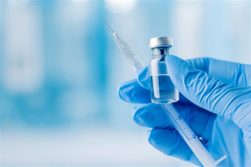
At Ultrasound Guided Injection LONDON, we are dedicated to providing expert care through cutting-edge ultrasound-guided injection treatments. Our clinic, based in London, specializes in precise, minimally invasive procedures to help patients manage pain and regain mobility. Whether you are struggling with joint pain, sports injuries, or chronic musculoskeletal conditions, we are here to provide advanced and effective solutions tailored to your needs.
Our Services
We offer a variety of ultrasound-guided injection therapies designed to provide targeted relief for a range of conditions. Our key services include:


Take control of your health with expert care from Ultrasound Guided Injection London – where precision meets relief!

Why Choose Ultrasound Guided Injection London?
At Ultrasound Guided Injection London, we believe that every patient deserves the highest standard of care. Here’s why people trust us:
Expert-Led Treatments
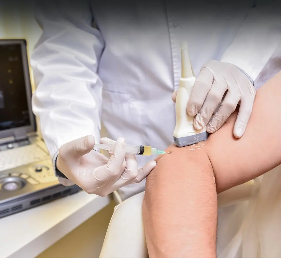
Our team of experienced specialists ensures accurate and targeted injections using state-of-the-art ultrasound technology.
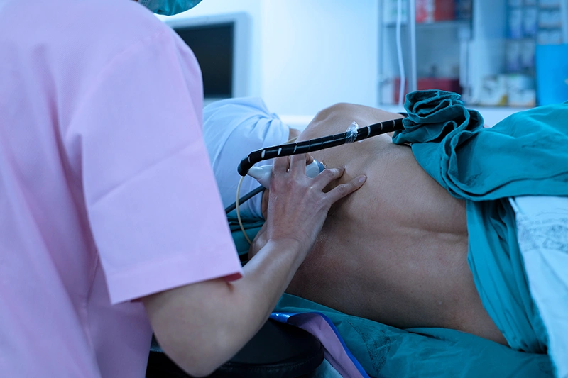
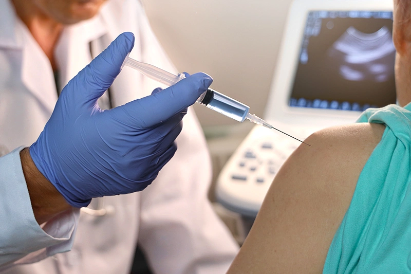
Advanced Imaging Techniques
Real-time ultrasound guidance improves precision, safety, and treatment effectiveness.
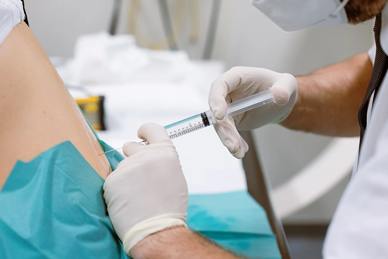
Minimally Invasive Approach
Our procedures involve minimal discomfort and reduced recovery times compared to traditional methods.
Personalized Treatment Plans
We understand that every patient is unique, and we tailor our treatments to address specific conditions and health goals.
Convenient London Location
Our clinic is easily accessible, making expert care available to those in need.
Proven Patient Outcomes
We focus on delivering effective pain relief and improved mobility to enhance the quality of life.
Joints We Treat
We specialize in treating pain and mobility issues in the following joints:

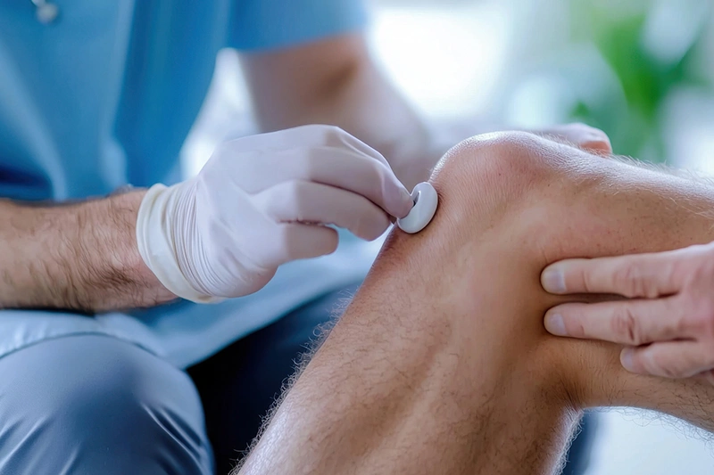
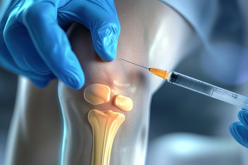
Knee
Commonly affected by arthritis, ligament injuries, and patellar tendonitis.

Elbow
Conditions such as tennis elbow and golfer's elbow can benefit from targeted injections.
Foot & Ankle
Issues like plantar fasciitis, Achilles tendonitis, and ankle sprains are treated effectively.

Wrist & Hand
Carpal tunnel syndrome, arthritis, and tendonitis in the wrist and hand are common concerns.
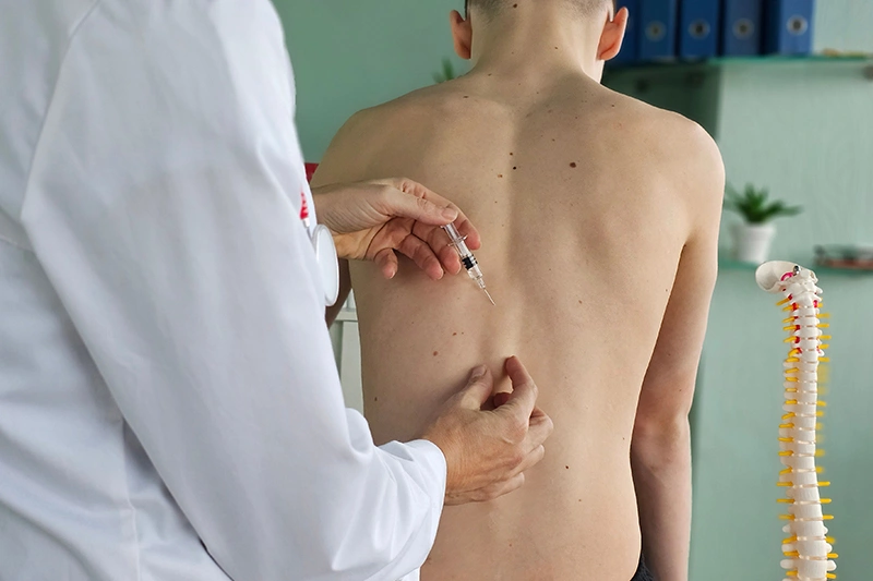
Spine
Nerve block injections help manage back pain, sciatica, and spinal arthritis.

Shoulder
Conditions like frozen shoulder, rotator cuff injuries, and bursitis can be relieved with guided injections.

Hip & Pelvis
Hip osteoarthritis, bursitis, and pelvic pain are treated with ultrasound-guided precision.
Conditions We Treat
At Ultrasound Guided Injection UK, we provide expert care for a variety of conditions, including:

Arthritis
Degenerative joint conditions causing pain, stiffness, and swelling.

Tendonitis
Inflammation of tendons, often due to repetitive motion or overuse.

Bursitis
Painful swelling in the fluid-filled sacs that cushion joints.

Frozen Shoulder
Stiffness and limited movement in the shoulder joint.

Carpal Tunnel Syndrome
Nerve compression in the wrist leading to numbness and pain.

Sports Injuries
Acute or chronic injuries affecting muscles, ligaments, or tendons.
Each condition is carefully assessed by our specialists to determine the best treatment plan for optimal results.
Don’t let pain control your life. Reach out to us today to schedule a consultation and take the first step toward pain-free living.
Frequently Asked Questions (FAQ)
Our treatments are effective for joint pain, arthritis, tendonitis, bursitis, frozen shoulder, carpal tunnel syndrome, and sports-related injuries.
The procedure is minimally invasive and well-tolerated. Most patients experience only mild discomfort.
Typically, the entire process takes 15-30 minutes, depending on the type of injection.
Pain relief can be experienced within a few days, but full effects may take up to two weeks.
Results vary based on the treatment type and individual response. Corticosteroid injections last weeks to months, while PRP and hyaluronic acid injections may provide longer-term benefits.